What is ABUS?
ABUS stands for Automated Breast Ultrasound. Ultrasound is a technology that creates images using reflected sound waves and has been used for decades in breast imaging. While not as sensitive as mammography, ultrasound provides some advantages that are helpful to supplement mammography screening. Ultrasound is superior to mammography in determining whether a mass is fluid-filled or solid. Ultrasound can detect masses that might be obscured on mammography due to dense breast tissue. And ultrasound does not use ionizing radiation to generate images like mammography.
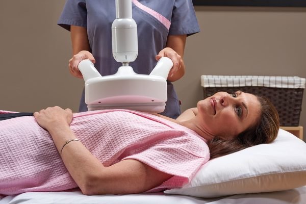
Automated Breast Ultrasound primarily differs from traditional or “hand-held” breast ultrasound in the size of the transducer used to acquire the images of the breast and in the way the images are displayed to the radiologist evaluating the images for breast cancer. The ABUS transducer is over three times longer than the transducers used in traditional breast ultrasound. With two to three sweeps of this larger transducer over the breast, advanced computer techniques reconstruct 3D-generated images of the entire breast. In contrast, traditional breast 2D ultrasound provides a small window in the breast and is used to evaluate specific areas, usually a focal palpable lump or a specific abnormality identified on mammography.
When the ABUS images are reconstructed and displayed, the radiologist has the opportunity to look at the entire breast, not just the small limited window. The development of automated technology that allows 3D images of the entire breast to be created, stored, and compared over time now allows ultrasound to be confidently used as a screening exam to supplement mammography.
How does it work?
Patients lie down on the exam table during an ABUS exam. Lotion is applied to the breast, and then the scanner is positioned to acquire the images. The entire exam takes approximately 30 minutes.
How do I know if ABUS is right for me?
ABUS is most effective in patients with dense breast tissue. Breast cancers are white on mammography and can sometimes be hidden by dense breast tissue, which is also white. Dense tissue is also white on ultrasound, but cancers are usually black or grey. Therefore, some cancers hidden on mammography are much more visible with ultrasound.
Breast density is determined by the radiologist who reads your mammogram, and the classification is always provided in the radiology report. Your doctor will inform you if your breasts are determined to be dense and if a supplemental exam is likely to be helpful.
What do I need to know about breast density?
Anatomically, breasts contain functional tissue associated with milk production mixed with differing amounts of fatty tissue. The breast tissue associated with milk production is white on mammography, while fatty tissue is gray. When there is enough white tissue on the mammogram to significantly camouflage a developing breast cancer, then the breast is considered dense.One concern is that dense breast tissue could hide a developing cancer.
In addition, relatively recent studies have demonstrated that dense breast tissue is associated with an increased risk of developing breast cancer. Women with extremely dense breast tissue have a 4x to 6x greater risk of developing breast cancer than women with fatty breast tissue.
How is ABUS different from a regular mammography?
While ABUS may detect some cancers that are hidden on mammography, mammography remains the most sensitive screening tool available for detecting breast cancers and saving lives. It is important to understand that mammography can detect some small breast cancers that cannot be seen on ultrasound – even in dense tissue. ABUS is most helpful as a supplement to mammography when breast tissue is known to be dense.
ABUS differs from mammography in that the breasts are not compressed between paddles during the exam. In addition, ABUS does not use ionizing radiation.
What should I expect from an ABUS screening?
Just like mammography and other types of breast ultrasound, not everything identified as a “lesion” on ABUS is a cancer. After an ABUS screening exam, you may be asked to come back for further evaluation, which usually includes a traditional breast ultrasound. While the smaller transducers of traditional breast ultrasound provide evaluation of a smaller area of tissue, these images can provide additional information not available on ABUS, such as higher definition images and evaluation of blood flow. This additional information can help determine if an identified abnormality is suspicious or not.


 Close
Close